Kijan Kolekstèktomi laparoskopik ye
Dec 08, 2021
Kolekstèktomi laparoskopik te vin tounen yon teknik chirijikal ki gen matirite, ki se aksepte pa majorite nan pasyan ki gen karakteristik sa yo nan mwens chòk, mwens doulè ak rekiperasyon rapid.
(1) Endikasyon
① Symptomatic gallstones.
② Symptomatic chronic cholecystitis.
③ Gallstone with diameter >3cm.
④ Filled gallstones.
⑤ Symptomatic and surgically indicated protuberant lesions of the gallbladder.
⑥ The symptoms of acute cholecystitis were relieved after treatment, and there were surgical indications.
⑦ It is estimated that the patient is well tolerated.
(2) Kontr relatif
① Acute attack of calculous cholecystitis.
② Chronic atrophic calculous cholecystitis.
③ Secondary choledocholithiasis.
④ History of upper abdominal surgery.
⑤ Fat body.
⑥ External abdominal hernia.
(3) Kontr absoli
① Acute cholecystitis with serious complications, such as gallbladder empyema, gangrene, perforation, etc.
② Gallstone acute pancreatitis.
③ With acute cholangitis.
④ Primary common bile duct stones and intrahepatic bile duct stones.
⑤ Obstructive jaundice.
⑥ Gallbladder cancer.
⑦ Protuberant lesions of the gallbladder are suspected to be cancerous.
⑧ Cirrhosis and portal hypertension.
⑨ Middle and late pregnancy.
⑩ Abdominal infection, peritonitis.
Chronic atrophic cholecystitis, gallbladder less than 4.5cm × 1.5cm, wall thickness >0.5cm (mesi à).
Akonpaye pa maladi emoraji ak malfonksyònman coagulation.
Moun ki gen fonksyon enkonplè nan ògàn enpòtan, ki difisil pou tolere operasyon ak anestezi, ak moun ki gen pesmekè kadyak (elektwokoagulasyon ak elektwokaterizasyon yo entèdi).
Kondisyon jeneral la se pòv, li pa apwopriye pou operasyon oswa pasyan an fin vye granmoun, pa gen okenn endikasyon fò nan kolekstèktomi, èrni dyafragmatik.
Sijè ki abòde lan endikasyon pou operasyon laparoskopik ap elaji ak devlopman nan teknoloji. Gen kèk maladi ki te orijinèlman kontr nan operasyon yo te eseye tou ranpli pa laparoskopi. Si choledocholithiasis segondè a te pasyèlman rezoud pa operasyon laparoskopik. Apre ou fin jwenn eksperyans ki nesesè yo, plis maladi ka trete pa operasyon laparoskopik.
(4) Pwosedi chirijikal
① Create pneumoperitoneum. Make an arc incision along the lower edge of the umbilical fossa, about 10mm long. If the lower abdomen has been operated on, cut the skin on the upper edge of the umbilical fossa to avoid the original surgical scar.
Operatè a ak premye asistan an chak kenbe pens sèvyèt twal pou leve miray vant la soti nan tou de bò fosa lonbrit la. Operatè a te kenbe zegwi pneumoperitoneum (Veress zegwi) ak gwo pous la ak dwèt endèks nan men dwat li, egzèse fòs sou ponyèt li, ak kout kouto nan kavite nan vant vètikal oswa yon ti kras oblik nan kavite basen an.
Nan pwosesis la nan twou, lè zegwi a kraze nan fascia a ak peritoneum, gen yon sans de zouti de fwa; Jije si pwent zegwi a antre nan kavite nan vant. Yon sereng ak saline nòmal ka konekte. Lè pwent zegwi a nan kavite nan vant, li montre presyon negatif. Konekte machin pneumoperitoneum la. Si presyon enflasyon an pa depase 1.73kpa, li endike ke zegwi pneumoperitoneum la nan kavite nan vant. Pa gonfle twò vit nan kòmansman an. Sèvi ak enflasyon koule ki ba, 1 2L pou chak minit.
At the same time, observe the intraperitoneal pressure on the pneumoperitoneum machine. The pressure during inflation should not exceed 1.73kpa. If it is too high, it indicates that the position of the pneumoperitoneum needle is incorrect, the anesthesia is too shallow and the muscle is not loose enough. Appropriate adjustment should be made. When the abdomen begins to bulge and the liver dullness boundary disappears, it can be changed to high flow automatic inflation until the predetermined value (1.73 2.00kpa) is reached. At this time, the inflation is 3 4L, the patient's abdomen is completely bulged, and the operation can be started.
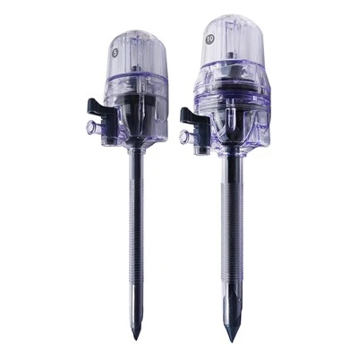
Lift the abdominal wall with towel pliers at the umbilical pneumoperitoneum needle and puncture with 10mm trocar. The first puncture has a certain "blindness", which is a more dangerous step in laparoscopy. Be extra careful. Rotate the trocar slowly and enter the needle evenly. When entering the abdominal cavity, there is a feeling that the resistance disappears suddenly. Open the closed air valve and gas escapes. This is the success of puncture. Connect the pneumoperitoneum machine to maintain constant pressure in the abdominal cavity. Then put the laparoscope in and puncture at each point under the monitoring of the laparoscope.
Anjeneral, twou 2cm anba pwosesis xiphoid la epi mete bwat 10mm pou zen egzeyat, aplikatè kranpon ak lòt enstriman; Pike 2cm anba kwen kostè nan liy dwat mitan klavikulèr la oswa 2cm anba rebò rektis abdominis ak kwen kostal devan axilè a ak 5mm trokar respektivman pou mete nan irigatè a ak gallbladder fiks atrab forceps. Nan moman sa a, pneumoperitoneum atifisyèl ak preparasyon yo te fini.
Akòz fabrike pneumoperitoneum ak premye twou trocar, gwo veso sangen ak trip nan kavite nan vant ka aksidantèlman blese, epi li pa fasil pou jwenn pandan operasyon an. Dènyèman, anpil moun te fè yon ti ouvèti nan lonbrit la pou jwenn peritoneum la epi dirèkteman mete trokar a nan kavite nan vant pou enflasyon. Apre fabrike siksè nan pneumoperitoneum, operasyon an te kòmanse.
② Dissect the Calot triangle. Grasp the neck of gallbladder or Hartmann's bursa with grasping forceps and traction to the upper right. It is best to draw the cystic duct perpendicular to the common bile duct in order to clearly distinguish the two, but pay attention not to draw the common bile duct into an angle. The serous membrane on the cystic duct was cut with an electrocoagulation hook, the cystic duct and cystic artery were passively separated, and the common bile duct and common hepatic duct were distinguished. Since it is close to the common bile duct, electrocoagulation should be used as little as possible to avoid accidental injury to the common bile duct. Use the electrocoagulation hook to separate the cystic duct upstream and downstream, and see the relationship between the cystic duct and the common bile duct. Place the titanium clip as close to the gallbladder neck as possible. There should be sufficient distance between the two titanium clips. The titanium clip should be at least 0.5cm away from the common bile duct. Cut between the two titanium clips with scissors, and do not use electric cutting or electrocoagulation to prevent damage to the common bile duct due to heat conduction. Then find the cystic artery behind it and cut it with titanium clip. After cutting off the gallbladder artery, do not pull hard to avoid breaking the gallbladder artery, and pay attention to the posterior branch of the gallbladder. Carefully peel off the gallbladder, electrocoagulation or hemostasis with titanium clip.
③ Cholecystectomy. Clamp the gallbladder neck and pull it upward, carefully peel it off along the gallbladder wall, and the assistant should assist in pulling to make the gallbladder and liver bed have a certain tension. Completely peel off the gallbladder and place it on the upper right side of the liver. The liver bed was hemostatic by electrocoagulation, carefully rinsed with normal saline, and checked for bleeding and bile leakage (a piece of gauze was disposed at the hepatic hilum, and checked for bile staining after removal). After absorbing all the water in the abdominal cavity, transfer the laparoscope to the lower sleeve of the xiphoid process and give way to the umbilical incision, so that the gallbladder containing stones greater than 1cm can be taken out from the umbilical incision with loose structure and easy expansion. If the stones are small, they can also be taken out from the puncture hole under the xiphoid process.
④ Remove the gallbladder. Put the toothed claw forceps into the abdominal cavity from the cannula at the umbilicus, grasp the residual end of the cystic duct under monitoring, slowly drag the gallbladder into the cannula sheath and pull it out together with the cannula sheath. When grasping the gallbladder, pay attention to placing the gallbladder on the liver to avoid accidental injury to the intestinal canal by sharp forceps. If the stone is large or the tension of the gallbladder is high, do not pull it out with force to avoid rupture of the gallbladder and leakage of stones and bile into the abdominal cavity. At this time, the incision can be enlarged with vascular forceps and taken out, or the incision can be expanded to 2.0cm with an expander. If the stone is too large, the incision can be extended. If bile leaks into the abdominal cavity, wet gauze shall be used to enter from the umbilical incision to suck up the bile.
Si wòch la twò gwo pou yo retire nan ensizyon an, ou ka tou louvri vezikulèr la an premye, souse kòlè a nan vezikulèr la ak yon aspiratè, epi retire li youn pa youn apre yo fin kraze wòch la ak forceps. Si yo jwenn yon wòch tonbe nan kavite nan vant, retire li. Apre w fin tcheke pa gen san ak likid nan kavite nan vant, rale soti laparoskop la, louvri valv la nan kanul la pou egzeyate gaz gaz kabonik nan kavite nan vant, ak Lè sa a, rale soti kanul la. Ensizyon an ak kanul 10mm sutured ak fil mens kòm kouch fascia pou 1 2 pwen, epi chak ensizyon fèmen ak fim adezif esteril.
(5) Gwo konplikasyon
① Bile duct injury. Bile duct injury is one of the most common and serious complications of laparoscopic cholecystectomy.
Ensidans nan blesi nan kanal bile ak flit kòlè se apeprè 10 pousan. Li ta dwe peye ase atansyon. Li se sitou akòz anatomi ki pa klè nan triyang Calot, espesyalman mank de vijilans kont varyasyon an komen nan kanal kòlè komen oswa kanal sistik. Lè separe kanal la sistik, kanal la kòlè te domaje tèmik inadvèrtans, pa te gen okenn flit kòlè pandan operasyon an, ak necrosis la ak tonbe nan tisi a nan zòn nan domaje tèmik apre operasyon an te kapab lakòz tou flit kòlè. Anplis de sa, gen souvan gwo kanal bile vagal nan kabann nan vezikulèr. Electrocoagulation intraoperative pa ka konplètman coagulation, ak flit kòlè kapab tou fòme. Manifestasyon prensipal yo nan blesi nan kanal bile yo se gwo doulè nan vant anwo, gwo lafyèv ak lajònis. Pasyan ki gen manifestasyon tipik yo anjeneral trete nan tan apre operasyon; Sepandan, kèk pasyan sèlman te montre tansyon nan vant, mank de apeti, lafyèv ki ba ak vin pi grav. Pasyan sa yo ta dwe byen obsève. Yo te rapòte ke yo te jwenn akimilasyon kòlè nan vant nan kèk mwa apre operasyon an. Pou jije si gen flit kòlè sitou depann sou ultrason oswa CT, ak Lè sa a, konfime pa pike zegwi amann anba pedagojik la nan ultrason oswa CT oswa epatocholangiography radyonukleid.
② Vascular injury. One is massive hemorrhage caused by needle tip injury to abdominal aorta, iliac artery or mesenteric vessels during pneumoperitoneum and trocar placement. There are many reports of death caused by trocar puncture. Therefore, after successful pneumoperitoneum, laparoscopy should peep the whole abdomen once to prevent missing vascular injury.
Lòt la se anatomi ki pa klè nan pòtal la epatik oswa move blocage nan atè epatik dwat la oswa atè epatik apwopriye akòz senyen nan atè vezikulèr. Genyen tou rapò sou blesi venn pòtal pandan anatomi. Te gen rapò sou necrosis epatik dwat ki te koze pa move blocage nan atè epatik.
③ Intestinal injury. Intestinal injuries are mostly accidental injuries caused by electrocoagulation, mainly because the electrocoagulation hook is not placed in the TV monitoring picture and is not found. Abdominal pain, abdominal distention and fever occur after operation, resulting in serious peritonitis, and its mortality is high.
④ Postoperative intraperitoneal hemorrhage. Postoperative intraperitoneal hemorrhage is also one of the serious complications of laparoscopic surgery. The injured parts are mainly the blood vessels near the gallbladder, such as hepatic artery, portal vein and abdominal aorta or vena cava during periumbilical puncture. The manifestations were hemorrhagic shock, abdominal bulge and peripheral circulatory failure. Open surgery should be performed immediately to stop bleeding.
⑤ Subcutaneous emphysema. The causes of subcutaneous emphysema are as follows: first, when making pneumoperitoneum, the pneumoperitoneum needle did not penetrate the abdominal wall, and high-pressure carbon dioxide entered the subcutaneous; Second, due to the small skin incision, the trocar is embedded very tightly, and the puncture hole of the peritoneum is relatively loose. During the operation, carbon dioxide gas leaks into the lower skin layer of the abdominal wall. Postoperative examination can find abdominal subcutaneous twisting pronunciation, generally without special treatment.
⑥ Others. Such as incisional hernia, incisional infection and abdominal abscess.